


Understanding Skin and Subcutaneous Lesions
Skin lesions can vary in size, shape and colour and range from benign lesions, such as skin tags and seborrhoeic keratoses, to pre-cancerous lesions. The latter include actinic keratoses and Bowen’s disease, which can progress to squamous cell carcinomas. Not all moles are simple freckles and some types are associated with an increased risk of melanoma.
Subcutaneous lesions lie beneath the skin and include sebaceous cysts and lipomas. These lesions are benign but may increase in size over time and can become troublesome.


Your Surgical Journey
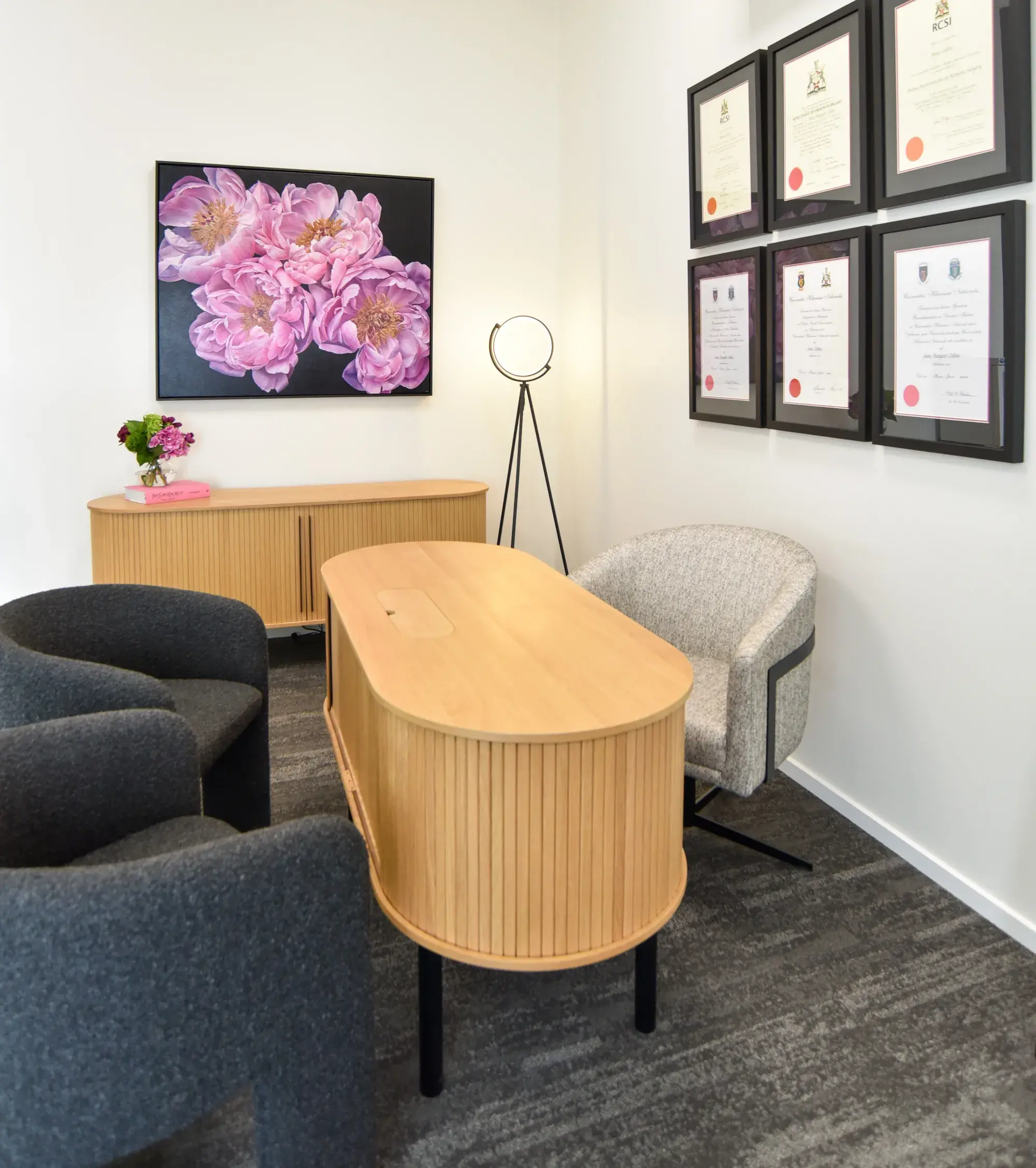
Your surgical journey begins with a comprehensive consultation with Dr Collins at The Surgery. This includes a full skin examination and, when appropriate, a review of your biopsy results or any relevant imaging.
Some lesions, such as seborrhoeic keratoses, can be treated with a simple shave excision. Other lesions, including pre-cancerous lesions and atypical moles, may require formal excision with a margin of surrounding skin. Subcutaneous lesions, such as cysts and lipomas, are typically excised. The aim is to remove the lesion while achieving an optimal cosmetic result.
Recovery and Aftercare
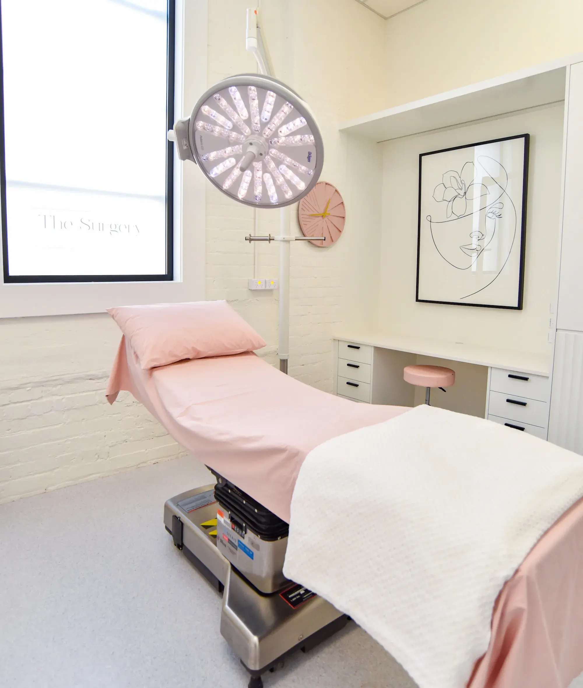
Recovery depends on the extent of the surgery. Smaller lesions are usually treated under local anaesthetic as a day procedure. More complex cases may require a general anaesthetic and a short hospital stay.
For desk-based roles, return to work is usually possible within a couple of days following procedures under local anaesthetic and within one to two weeks following general anaesthesia. More physically demanding roles may require a longer recovery period.
Management and Next Steps
Management is guided by the clinical features of the lesion and, where appropriate, histological findings.
A consultation with Dr Collins allows for a personalised assessment and discussion of your results and management plan. Ongoing skin checks are recommended, particularly if you have a history of skin cancer or significant sun exposure.
Skin and Soft Tissue Lesions in Dunedin with Dr Anne Collins

Frequently Asked Questions
Also known as an age spot, a seborrhoeic keratosis is a common benign lesion. It often appears as a well-defined, slightly raised lesion with a warty or “stuck-on” appearance. These lesions are harmless but can be removed if they become irritated or troublesome.
Actinic keratoses and Bowen’s disease are pre-cancerous skin lesions caused by cumulative sun exposure. Actinic keratoses typically appear as rough, scaly patches, while Bowen’s disease presents as a well-defined, red plaque. Both have the potential to progress to squamous cell carcinoma and are usually treated to reduce this risk.
Certain moles, known as atypical or dysplastic moles, are associated with an increased risk of melanoma. These moles may appear larger than usual, with irregular borders, variation in colour or an uneven shape. If a mole changes in size, shape or colour, it should be assessed promptly.
An epidermal cyst is a benign lesion that forms beneath the skin and often contains keratin, sometimes with a visible central punctum. A lipoma is a benign growth of fatty tissue that typically feels soft, mobile and sits deeper beneath the skin. Both are harmless but may be removed if they increase in size or become uncomfortable.
The surgical technique depends on the type of lesion. Some lesions can be treated with a shave excision, while others require formal excision with an appropriate margin of surrounding skin.
A shave excision involves removing a lesion from the surface of the skin using a fine surgical blade. It is commonly used for benign lesions and does not require stitches.
Certain benign and pre-cancerous lesions can be treated with topical therapies, including creams or cryotherapy. These options will be discussed with you where appropriate.
If your lesion is located beneath the skin, an ultrasound may be arranged in the first instance to help confirm the diagnosis and assist with surgical planning. In some cases, further imaging such as an MRI may be required if additional detail is needed.
Recovery depends on the extent of the surgery. Shave excisions and excisional biopsies are usually carried out under local anaesthetic as a day procedure. More complex cases may require a general anaesthetic and a short hospital stay. Specific aftercare instructions will be provided to support your recovery.
For desk-based roles, return to work is usually possible within a couple of days following procedures under local anaesthetic and within one to two weeks following general anaesthesia. More physically demanding roles may require a longer recovery period.
Driving is usually possible after one to two weeks, once you can safely control the vehicle and perform emergency manoeuvres comfortably.
The appearance of the scar depends on the type of procedure performed. Shave excisions usually leave a flat, pale mark, while formal excisions result in a linear scar that fades over time. The aim is to remove the lesion while achieving an optimal cosmetic result.
Lesions that are biopsied or excised are routinely sent for histological analysis and a formal report is generated. This does not apply to superficial treatments, such as topical therapies or cryotherapy, where tissue is not removed for analysis.
If a lesion is found to be pre-cancerous or malignant, further treatment or follow-up may be required. This will be discussed with you in detail and a personalised management plan will be tailored to your diagnosis.

Online Enquiry
We would love to hear from you. If you have any questions or would like to make an appointment, please complete your details below.